MitochondriaAnimals

Elamipretide improves oocyte quality and restores fertility in aged mice

The peptide also boosted maturation and fertilization in aged human oocytes by activating the vitamin B6 and VEGF pathway.

Aging Cell

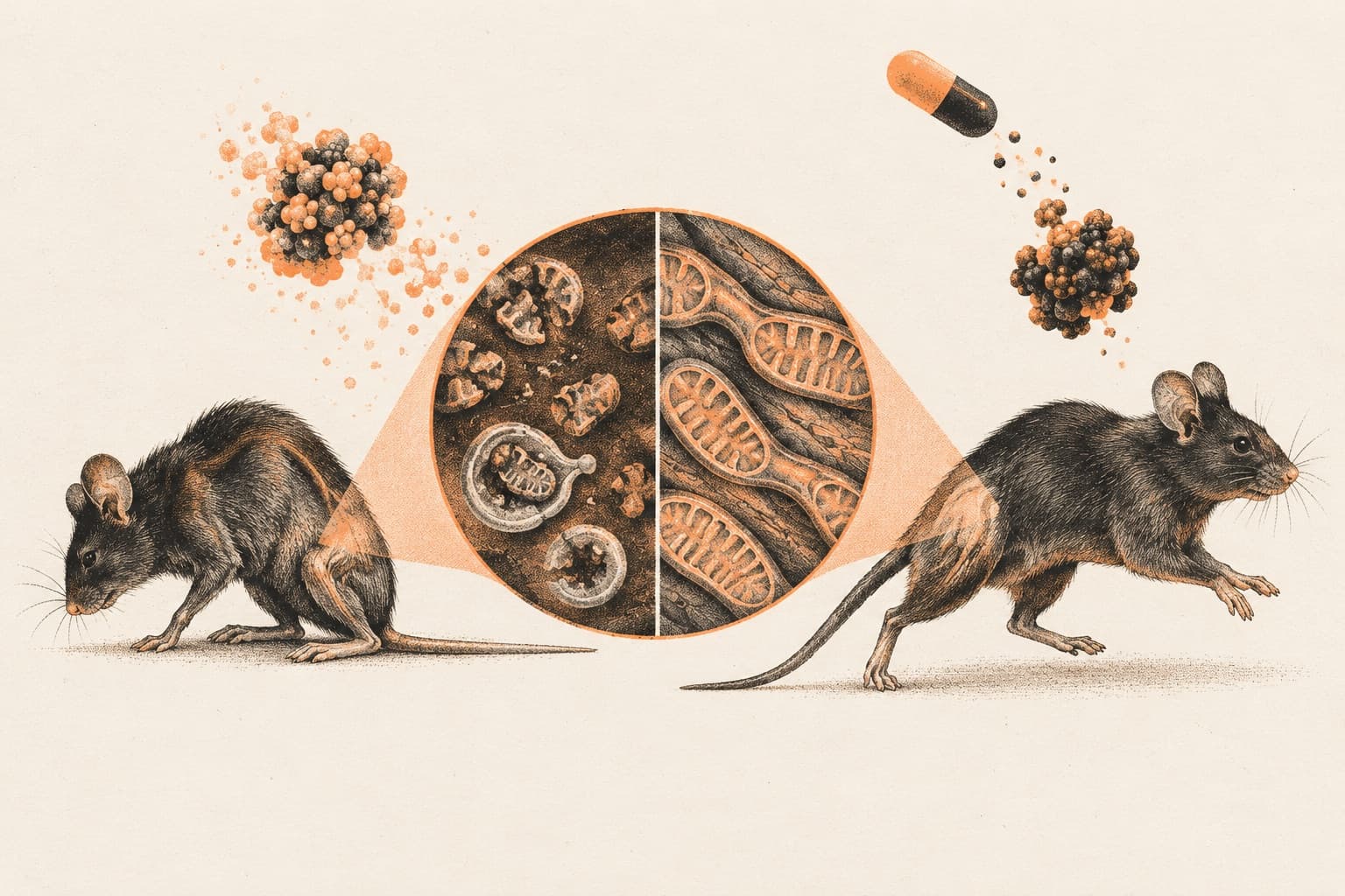
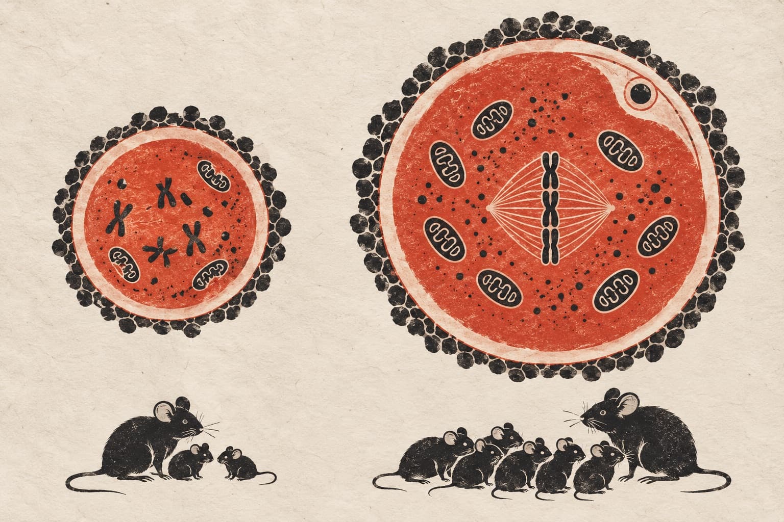
In female mice experiencing reproductive aging, administration of the peptide elamipretide increased litter size and ameliorated age-related declines in oocyte quality. Researchers observed that the treatment improved both nuclear and cytoplasmic maturation in aged mouse oocytes. Metabolomic and transcriptomic profiling revealed broad restoration of biological pathways, accompanied by enhanced mitochondrial metabolism, organelle reorganization, and cytoskeletal dynamics. Mechanistically, elamipretide alleviated age-associated maturation defects by synergistically activating the vitamin B6-VEGF signaling axis. Beyond rodent models, the authors evaluated elamipretide across other species. In aged human oocytes, the peptide significantly promoted maturation, fertilization, and cleavage rates. It also rescued the developmental competence of porcine oocytes subjected to oxidative damage.

Why it matters

Ovarian aging and declining oocyte quality are primary drivers of reproductive senescence and infertility. Identifying interventions that restore mitochondrial metabolism and cytoplasmic maturation provides key mechanistic targets for extending female reproductive longevity.

Caveats

The fertility and litter size benefits were demonstrated in mice, whereas effects on human oocytes were tested only in cultured cells. Clinical trials are necessary to verify whether elamipretide improves pregnancy and live birth outcomes in women.

The paper

Elamipretide Rejuvenates Oocyte Quality and Restores Female Fertility During Reproductive Aging

Zhang HL, Wang Y, Wang C et al.

Aging Cell · 1 Oct 2026
See all
Nature AgingAnimals

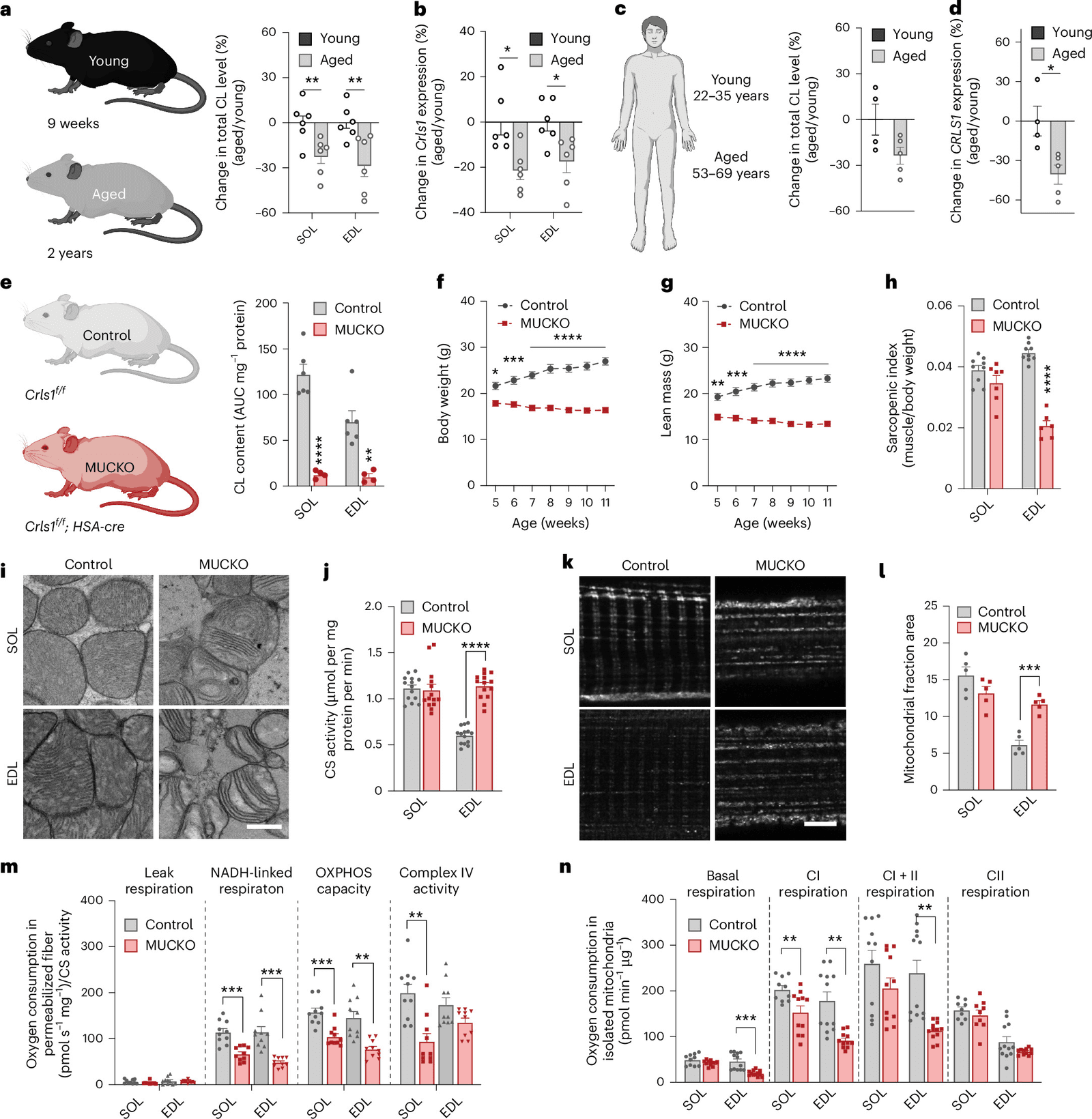
Cardiolipin loss drives muscle fiber shifts during aging via a nuclear receptor

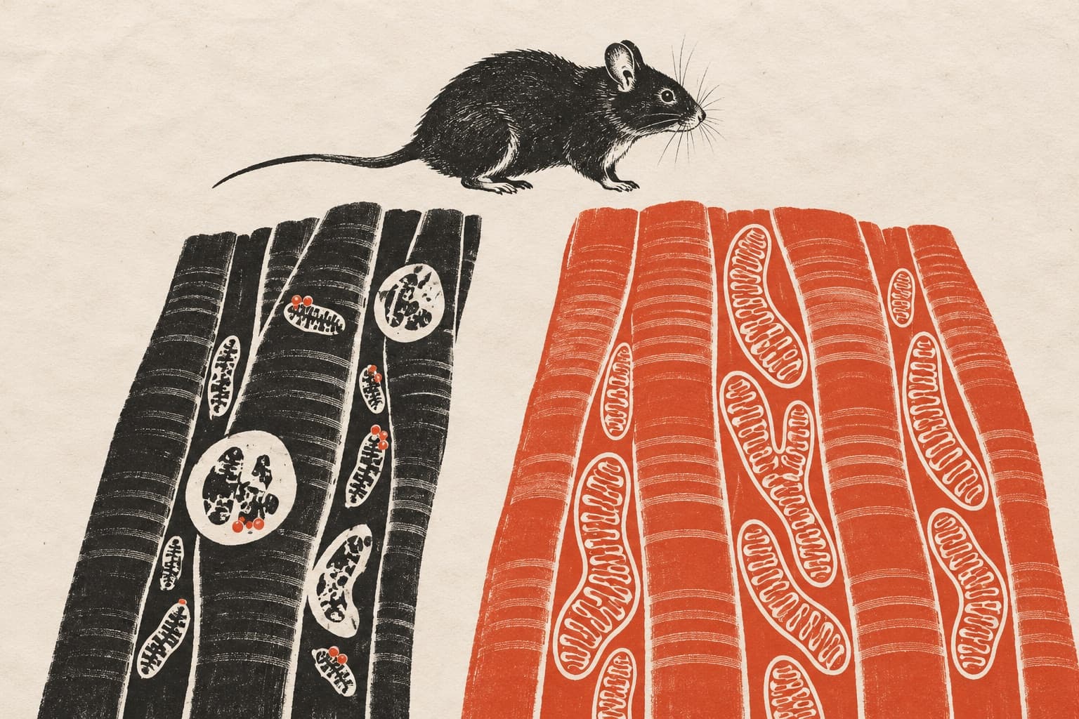
Restoring cardiolipin synthesis in knockout mice reversed muscle atrophy and prevented premature death.Restoring cardiolipin synthesis in knockout mice reversed muscle atrophy and prevented premature death. In mice and human skeletal muscle, the loss of the mitochondrial membrane lipid cardiolipin links organelle dysfunction to aging-related fiber adaptations.

Figure 1
bioRxivAnimalsPreprint

Early-life mitochondrial DNA mutations drive age-related pathology in mice

Manipulating mitochondrial fusion can alter tissue-specific selection against deleterious variants that arise early in development.Manipulating mitochondrial fusion can alter tissue-specific selection against deleterious variants that arise early in development. In a new preprint, researchers engineered mice to confine mitochondrial DNA mutagenesis to specific developmental windows.

bioRxivAnimalsPreprint

Inhibiting miR-128-3p restores muscle mass and function in aged mice

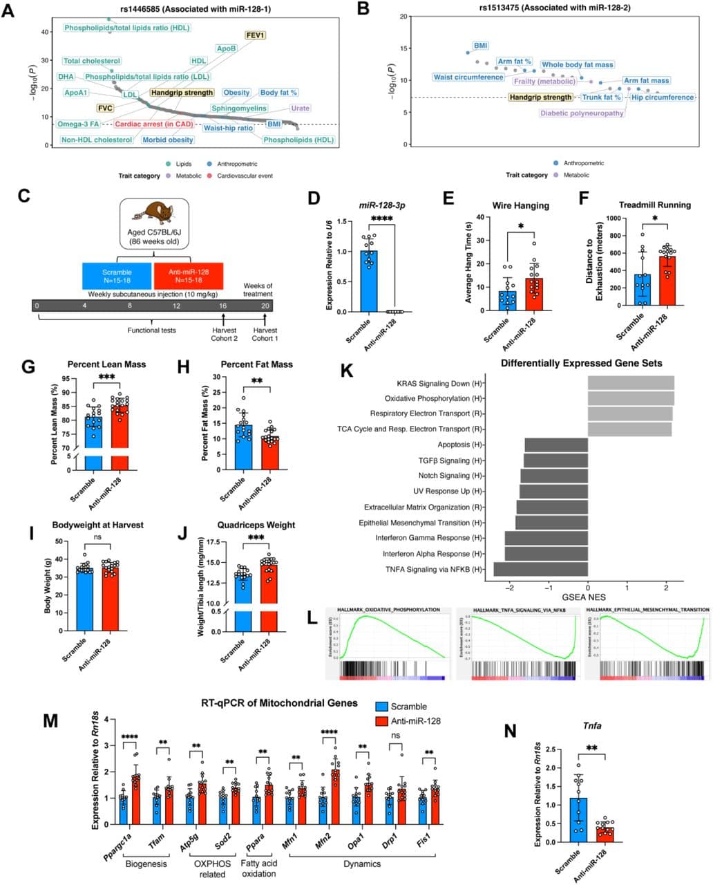
The microRNA inhibitor also improved cardiac outcomes after infarction and reduced pathology in mouse and pig models of muscular dystrophy.The microRNA inhibitor also improved cardiac outcomes after infarction and reduced pathology in mouse and pig models of muscular dystrophy. In aged mice, inhibiting the microRNA miR-128-3p with antisense oligonucleotides restored muscle mass and function, according to a recent preprint.

SNPs in the miR-128 loci are associated with muscle and pulmonary function in humans and anti-miR-128 improves muscle function and transcriptional programs in aged mice.
Aging CellAnimals

Prodh2 inhibition alleviates muscle atrophy and restores strength in COPD mice

TNF-alpha triggers a mitochondrial immune pathway via Prodh2 that damages myoblasts, while silencing the enzyme restores muscle strength in mice.TNF-alpha triggers a mitochondrial immune pathway via Prodh2 that damages myoblasts, while silencing the enzyme restores muscle strength in mice. In a study examining sarcopenic COPD patients and mouse models, researchers found that the mitochondrial enzyme Prodh2 mediates…

bioRxivAnimalsPreprint

Depp1 drives muscle loss under fasting and low oxygen in mice

The study reveals that the protein localizes to mitochondria to regulate autophagy and mitochondrial degradation during nutrient and oxygen…The study reveals that the protein localizes to mitochondria to regulate autophagy and mitochondrial degradation during nutrient and oxygen limitation. In a new preprint, researchers studied mice to determine how low oxygen and nutrient scarcity trigger skeletal muscle loss.