MitochondriaHumans

Darker skin pigmentation links to fewer mitochondrial DNA mutations from sun exposure

Melanin protects against mitochondrial genome damage and clonal expansion in chronically sun-exposed human skin.

Pigment Cell & Melanoma Research

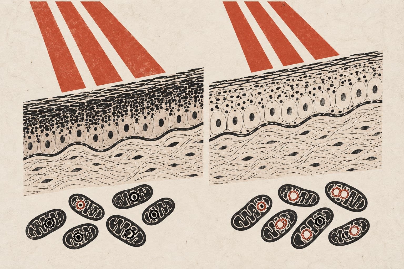
In an observational study of six older men, researchers analyzed how skin pigmentation influences mitochondrial DNA (mtDNA) stability under chronic sun exposure. The team performed targeted mitochondrial sequencing on paired skin biopsies from the photoexposed forearms and photoprotected buttocks of three Black African and three White Northern European participants. They identified 21 high-confidence somatic mtDNA variants across the samples. Photoexposed light skin possessed six-fold more variants than dark skin and was enriched for ultraviolet radiation signatures, such as C>T transitions and CC>TT substitutions. These variants in light skin altered multiple oxidative phosphorylation genes and frequently reached moderate-to-high heteroplasmy. Conversely, dark skin displayed a narrower variant profile dominated by a recurrent T414G substitution and lower overall heteroplasmy.

Why it matters

Mitochondrial genomic instability and respiratory chain impairment are hallmarks of tissue aging. Showing that melanin shields mitochondrial DNA helps explain why darker skin shows greater structural resistance to ultraviolet-induced photoaging.

Caveats

The findings are based on a very small sample size of six male donors and rely on an observational study design. Larger, more diverse cohorts are needed to confirm how broadly these findings apply across sexes and skin types.

The paper

Skin Pigmentation Associates With Reduced mtDNA Variant Burden in Photoexposed Skin

Mambwe B, Langton AK

Pigment Cell & Melanoma Research · 3 Oct 2026
See all
Nature AgingAnimals

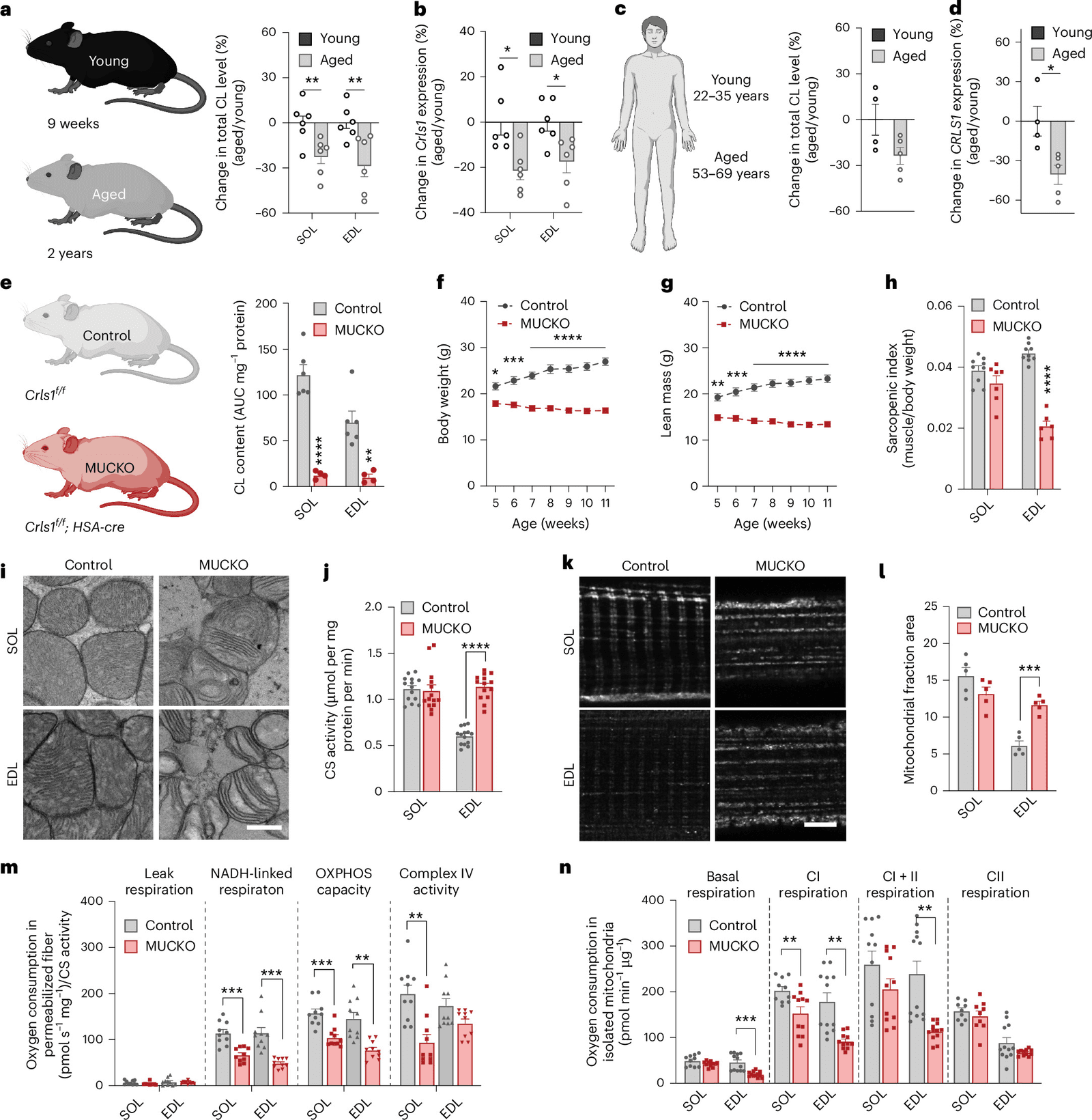
Cardiolipin loss drives muscle fiber shifts during aging via a nuclear receptor

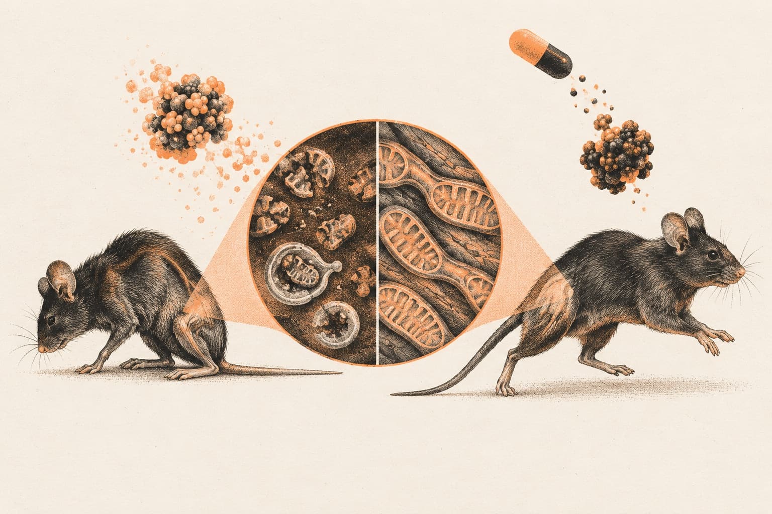
Restoring cardiolipin synthesis in knockout mice reversed muscle atrophy and prevented premature death.Restoring cardiolipin synthesis in knockout mice reversed muscle atrophy and prevented premature death. In mice and human skeletal muscle, the loss of the mitochondrial membrane lipid cardiolipin links organelle dysfunction to aging-related fiber adaptations.

Figure 1
bioRxivAnimalsPreprint

Early-life mitochondrial DNA mutations drive age-related pathology in mice

Manipulating mitochondrial fusion can alter tissue-specific selection against deleterious variants that arise early in development.Manipulating mitochondrial fusion can alter tissue-specific selection against deleterious variants that arise early in development. In a new preprint, researchers engineered mice to confine mitochondrial DNA mutagenesis to specific developmental windows.

bioRxivAnimalsPreprint

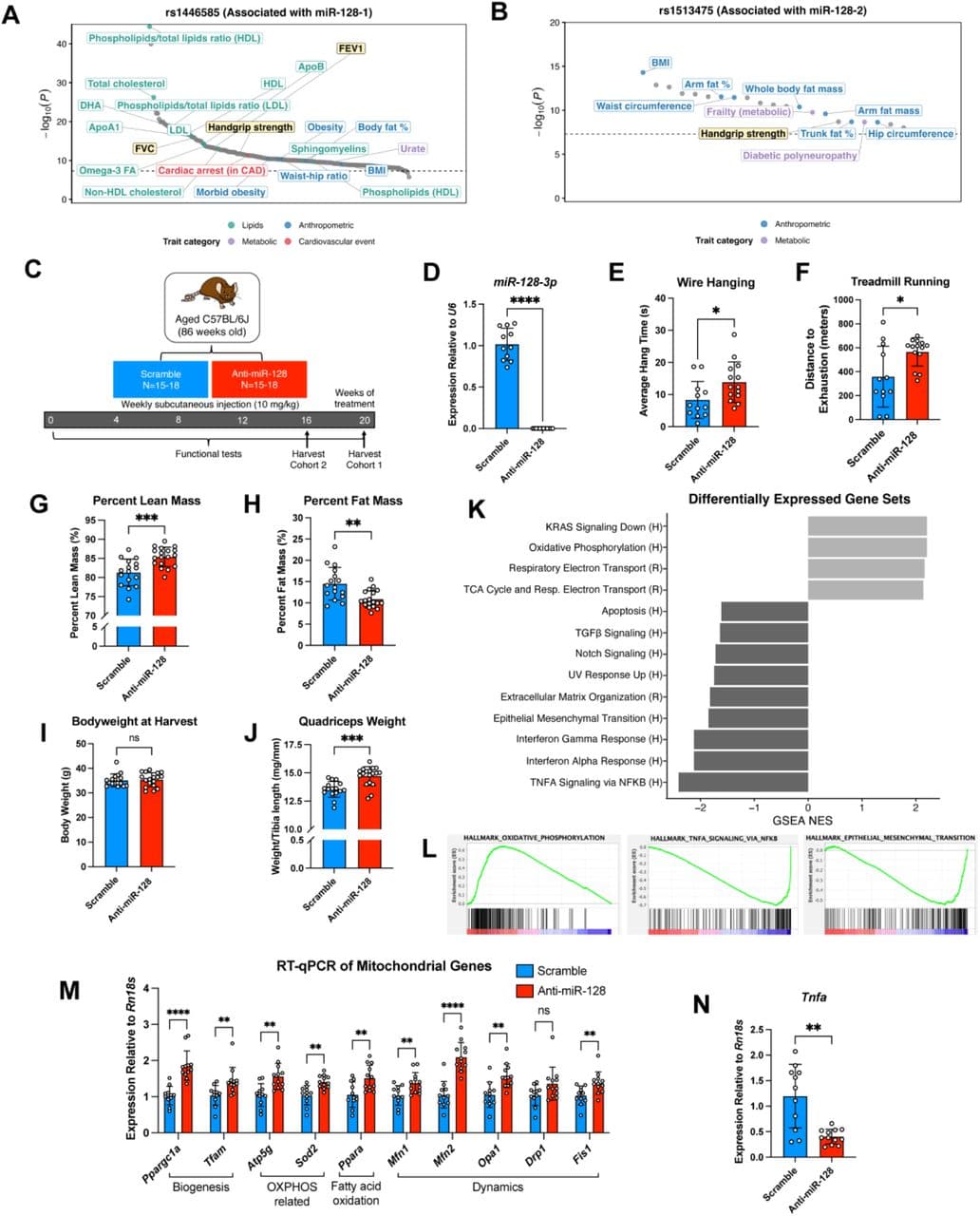
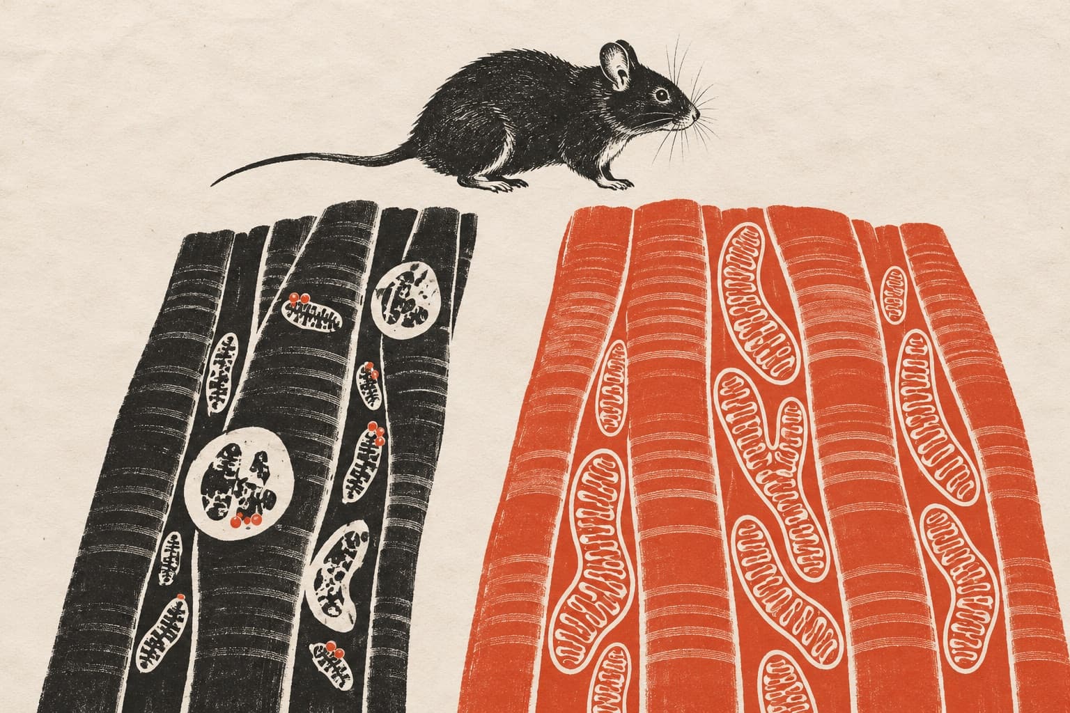
Inhibiting miR-128-3p restores muscle mass and function in aged mice

The microRNA inhibitor also improved cardiac outcomes after infarction and reduced pathology in mouse and pig models of muscular dystrophy.The microRNA inhibitor also improved cardiac outcomes after infarction and reduced pathology in mouse and pig models of muscular dystrophy. In aged mice, inhibiting the microRNA miR-128-3p with antisense oligonucleotides restored muscle mass and function, according to a recent preprint.

SNPs in the miR-128 loci are associated with muscle and pulmonary function in humans and anti-miR-128 improves muscle function and transcriptional programs in aged mice.
Aging CellAnimals

Prodh2 inhibition alleviates muscle atrophy and restores strength in COPD mice

TNF-alpha triggers a mitochondrial immune pathway via Prodh2 that damages myoblasts, while silencing the enzyme restores muscle strength in mice.TNF-alpha triggers a mitochondrial immune pathway via Prodh2 that damages myoblasts, while silencing the enzyme restores muscle strength in mice. In a study examining sarcopenic COPD patients and mouse models, researchers found that the mitochondrial enzyme Prodh2 mediates…

bioRxivAnimalsPreprint

Depp1 drives muscle loss under fasting and low oxygen in mice

The study reveals that the protein localizes to mitochondria to regulate autophagy and mitochondrial degradation during nutrient and oxygen…The study reveals that the protein localizes to mitochondria to regulate autophagy and mitochondrial degradation during nutrient and oxygen limitation. In a new preprint, researchers studied mice to determine how low oxygen and nutrient scarcity trigger skeletal muscle loss.